Fluorescence is a molecular phenomenon in which a substance absorbs light of one color and almost immediately emits light of another color, with lower energy and therefore 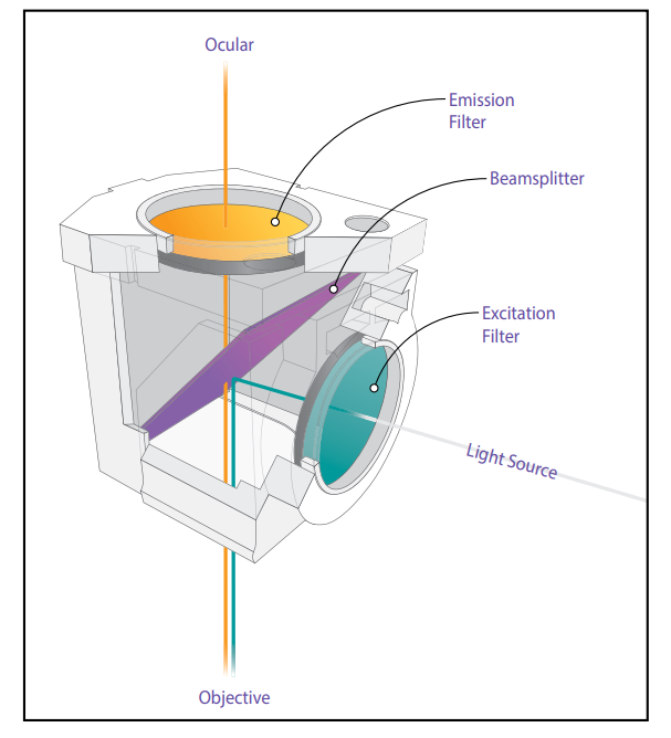 longer wavelength This process is known as excitation and diffusion. Many materials, both organic and inorganic, exhibit some fluorescence. In the early days of fluorescence microscopy (at the turn of the century) microscopes looked at this primary fluorescence, or autofluorescence, but now many dyes have been developed that fluoresce very brightly and are used to selectively stain sections of the sample. This method is called secondary or indirect fluorescence. These dyes are called fluorochromes, and when conjugated with other organic active substances such as antibodies and nucleic acids, they are called fluorescent probes or fluorophores. (These different terms are often used interchangeably.) There are now fluorochromes that have characteristic emission maxima in the near infrared as well as the blue, green, orange, and red colors of the spectrum. When indirect fluorescence through fluorochromes is used, the autofluorescence of a sample is generally considered undesirable: it is often the main source of unwanted light in the microscope image
These color spectra are quantitatively described by the wavelength of light. The most common unit of wavelength to describe the fluorescence spectrum is the nanometer (nm)
The colors of the visible spectrum can be divided into approximate wavelength values :
- violet and indigo 400 to 450 nm
- blue and blue blue 450 to 500 nm
- green 500 to 570 nm
- yellow and orange 570 to 610 nm near red 750 nm
Fluorescent filters
: Exciter Filters: Exciter filters allow certain wavelengths of the illuminant to pass through to the sample. They are designed to transmit only selected excitation wavelengths
Excitation filters are essential for exciting fluorophores in the sample
Barrier Filters: Barrier filters (also known as diffusion filters) suppress or block excitation wavelengths
They allow only selected emission wavelengths to pass to the eye or other detectors
Barrier filters help to separate the emitted fluorescence signal from the excitation light
Two-color rays (two-color mirrors)
Dichroic beams effectively reflect excitation wavelengths and allow emission wavelengths to pass through
They are placed after the driving filter but before the blocking filter
These specialized filters are oriented at an angle of 45 degrees to the paths of excitation and emission of light
Types of filters
Fluorescence filters were traditionally made of colored glass or gelatin sandwiched between glass
Today, interference filters (dielectric coating on glass) are more commonDriving filters are often of interference type and some barrier filters are also interference filters
Dichroic beams are specialized interference filters
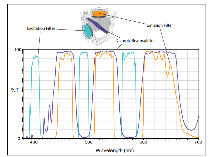
Filter curves (spectrum)
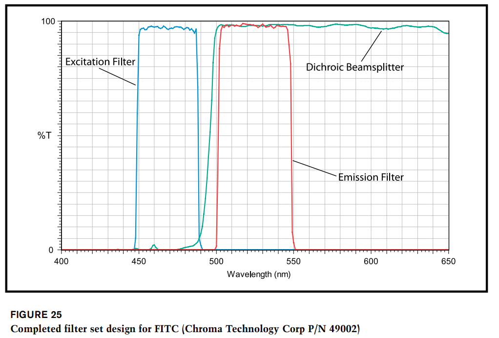
Filter curves show percent transmission (or logarithm of percent) versus wavelength
Drive filters labeled with letters (eg BP for bandpass) indicate their characteristics
Short pass (SP) and long pass (LP) filters can be combined to narrow the wavelength band
Numbers associated with filters may refer to the wavelength of maximum transmission or the 50% transmission point

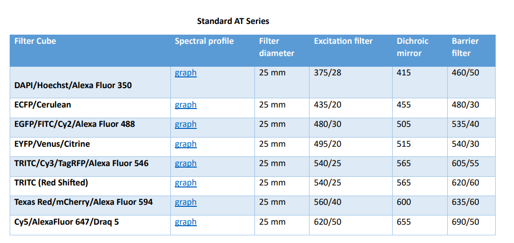
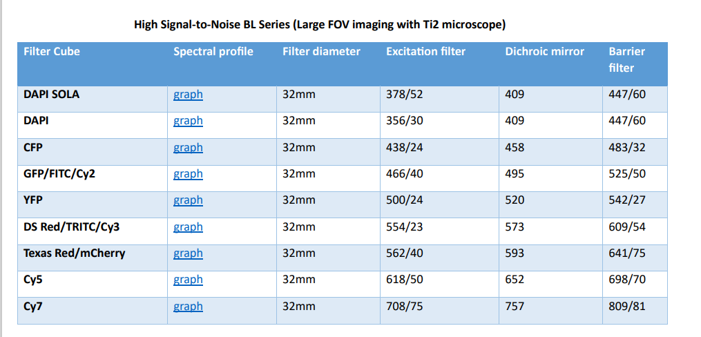
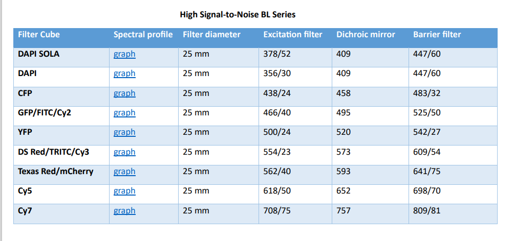

https://www.thorlabs.com/newgrouppage9.cfm?objectgroup_id=2990
Fluorescence imaging filters
دیدگاه خود را بنویسید