Blastomere fixation techniques
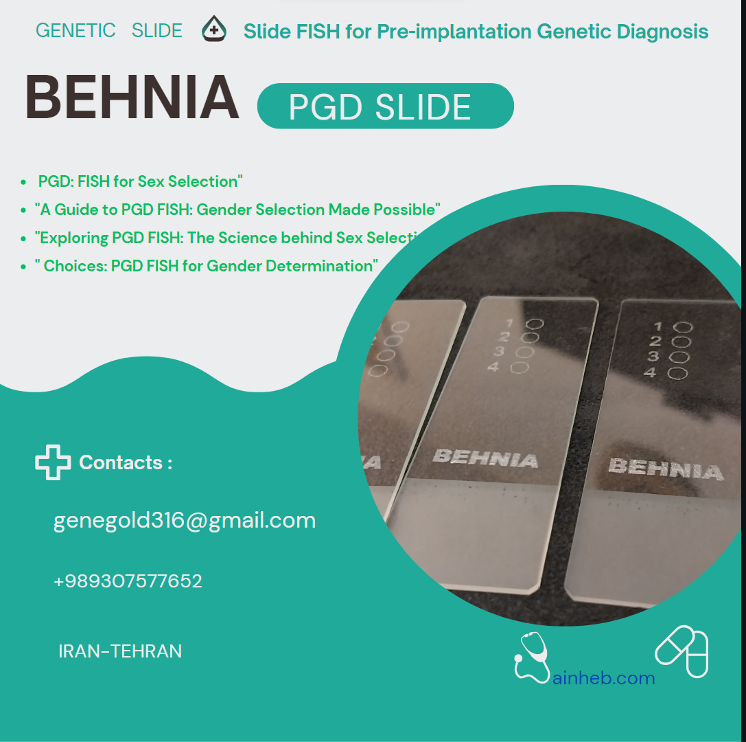
 اسلاید اختصاصی PGD
بررسی نمونههای FISH PGD زیر میکروسکوپ یکی از مهمترین و پرکاربردترین روشهایی تشخیصی در آزمایشگاهها است. این کار مستلزم تهیه لام به روش مناسبی است. از آنجایی که نمونهها متنوع هستند، روش تهیه لام از نمونه نیز متفاوت است.
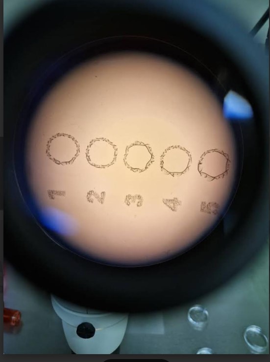
اسلاید های طراحی شده در شرکت ژن های طلایی مناسب این تکنیک بوده و برای مراکز ژنتیک مختلف دنیا صادر میشود. این اسلایدها به جنین شناسان کمک شایانی میکند برای قراردادن بلاستومر در محدوده مشخص شده و با خاصیت هیدروفیل که توسط مواد اختصاصی ژنهای طلایی طراحی شده حالت ابگریز و آبدوست داشته و بلاستومر در داخل چاهک می ماند.
این اسلایدهای نیاز به شتسشو ندارند و نیاز به نظافت با الکل یا مایع شوینده های که خطرناک برای جنین شناسی و بخش ژنتیک هست نیستند آماده برای قراردادن نمونه در روی اسلاید .
اسلایدهای به گونه ای طراحی شدند شما به راحتی با جنین شناس خود مختصات بلاستومر و نحوه کوردینت اصلی بلاستومر را شناسایی میکنید .

این لام های اختصاصی تکنیک PGD FISH میباشد .
1. چاهک های مناسب انتخاب شده تا میزان مصرف پروب مقرون به صرفه باشد.
2. به علت مناسب بودن فضای بلاستومر سریع پیدا میشود .
3.در یک ران کاری 4 بلاستومر از بیماران مختلف قابل بررسی میباشد
4.فاقد سیگنالهای کاذب میباشد به علت شستشوی اسلاید ها با ماده اختصاصی N .
5.در تهیه این لامها موارد مصرف تمامی مواد و پروب محاسبه شده و همچنین فاصله ها هم اندازه و شماره گذاری اختصاصی دارند تا قسمت انتخاب شده مناسب و مطمین باشد .
- ویژه آموزشی

FISH PGD Study

Preimplantation Genetic Testing FAQs
What are the risks, benefits, and limitations of PGT
What will happen with my embryos
What is the cost of testing
How do I prepare for my PGT appointment with a genetic counselor
How is PGT performed
What are the risks, benefits, and limitations of PGT
The risks, benefits, and limitations of PGT are reviewed in detail during a genetic counseling consult. It is important to understand these prior to completion of PGT in an IVF cycle. There are risks associated with the biopsy for PGT, as well as the freeze/thaw process. It is possible that there may be no embryos recommended for transfer based on the PGT results. PGT does not identify all genetic risks and does not replace screening and testing options that may be recommended during a pregnancy
What will happen with my embryos
After the CRH embryologist performs a biopsy to remove several cells for PGT, your embryo(s) will be frozen and retained in storage. The embryo biopsy sample(s) will be delivered via by a courier to the PGT laboratory for testing. Prior to cycle start, you will complete PGT consent and directive forms with a CRH staff member. In the directive form, you will elect whether you would prefer affected/abnormal embryos are donated to research or discarded from storage. If you complete both PGS and PGT-M, you will have the option to store all embryos with a normal PGS result regardless of the PGT-M result. Please speak with your genetic counselor or physician if you have questions or concerns prior to signing these documents
What is the cost of testing
The exact cost for PGT varies based on the type of testing completed and number of embryo samples tested, in addition to other factors such as potential insurance coverage and the laboratory at which testing will be completed. Your CRH genetic counselor can provide you with self-pay cost estimates or connect you with the billing department at the PGT laboratory, as appropriate Please note that the cost for PGT itself is separate to any associated fees from laboratorlaboratories, which may include the charges for procedures such as embryo biopsy and freeze
Please follow up with your financial navigator if you have any questions or concerns about expected costs prior to cycle start
Fluorescent in-situ hybridization (FISH)
is the technique that has traditionally been used in the study of chromosomal abnormalities
It only allows the analysis of certain regions of 9 chromosomes (13, 15, 16, 17, 18, 21, 22, X and Y). However, these chromosomes are involved in aneuploidies that can lead to repeated miscarriages or the birth of sick children
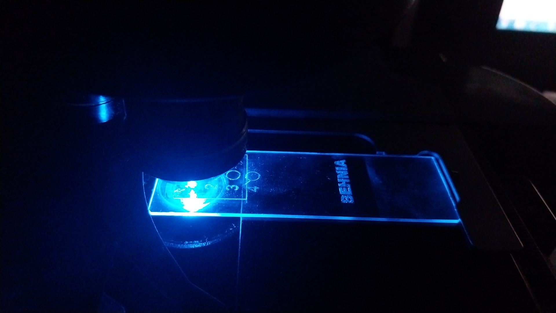
The process consists of adding fluorescent probes for specific regions of the chromosomes to be analyzed. It is then possible to visualize the fluorescent signal through a special microscope and to detect whether any of the analyzed regions are missing or, on the contrary, if there are more copies than they should be
آموزش مراحل تکنیک FISH PGD توسط تیم ما
09307577652

دیدگاه خود را بنویسید